
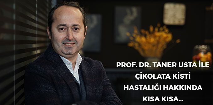
A Quick Look at Chocolate Cyst Disease with Prof. Dr. Taner Usta…
Endometriosis is seen in 1 in every 10 women of reproductive age (mostly between the ages of 15 and 49). Endometriosis is seen in approximately 176 million women worldwide.
Endometriosis is seen in 1 in every 10 women of reproductive age (mostly between the ages of 15 and 49). Worldwide, endometriosis is seen in approximately 176 million women. In our country too, we estimate that approximately 2 million of our women have endometriosis.
Endometriosis is a disease of the reproductive years and is seen most often in the thirties. It can also affect women’s physical, mental and social lives. Women’s lack of awareness or the normalisation of the symptoms lengthens the time that passes from the first symptoms of the disease being noticed to the diagnosis being made.
It is seen at a rate of 70% in women who have painful periods, painful intercourse and chronic groin pain. It is seen at a rate of 30-40% in women with a problem of infertility. In general it is seen in one in every 10 women of reproductive age (15-49 years). Although endometriosis is not commonly seen in the 13-19 age group, whom we call adolescents, in young girls who have severe menstrual pain and in whom no response to painkillers is observed, the frequency of endometriosis rises to as high as 70%.
It is the settling in organs outside the uterus of the tissue that lines the inside of the uterus and is called the endometrium. In order of frequency, the organs in which it is seen have been reported as the peritoneal membrane lining the inside of the abdomen, the ovaries, the tubes, the vagina, the bowel, the bladder, the ureter, the umbilicus (navel), the sites of stitches from previous operations, the lungs, the diaphragm, the nose, the brain, the eye, breast tissue, the heart and the spleen. Most frequently, at a rate of 60-75%, it is seen in the ovaries.
What are the symptoms of endometriosis?
The endometriosis foci that ultimately form grow and bleed according to the changes in the hormone oestrogen during the menstrual cycle, spread deeper and, as a result of this, create pain and adhesions in the areas where they are located. While the cells of the immune system try to eliminate the endometriosis foci, an inflammatory condition also develops alongside, and as a result of this, scar tissue and serious adhesions occur.
Painful periods, painful sexual intercourse (pain radiating to the groin and the anus), infertility, constipation, indigestion, bloating, abdominal pain, a feeling of discomfort in the pelvis, that is, in the lower part of the abdomen, menstrual irregularity, spotting before the period, frequent urination, blood in the urine and flank pain are the patient complaints that bring endometriosis to mind (Table 1). From time to time it causes no complaints at all and is seen during an operation or an ultrasound scan.
Table 1. The clinical complaints that frequently suggest endometriosis may be present.
First of all, as with every disease, the patient’s complaints and personal history are taken. Then a gynaecological examination and ultrasound scan are performed. During the pelvic examination, tenderness, an expression of pain and restricted movement of the uterus and ovaries suggest endometriosis. From time to time, during the speculum examination, blue-purple nodular lesions can be seen behind the cervix (the neck of the womb), and this simple finding indicates the presence of endometriosis in the deep tissues of the pelvis. For this reason, pelvic examination with a speculum is important in making the diagnosis.
The most informative of the auxiliary methods in diagnosis is ultrasonography. With ultrasonography, the chocolate cysts in the ovary that we call endometriomas, serious adhesions between the ovaries and the uterus and, from time to time, endometriotic nodules in the bowel, the rectum and the bladder can be visualised. The advanced imaging methods MRI and CT are useful especially in situations where endometriosis involves the deep pelvic tissues, which we call deep endometriosis.
Another auxiliary method is blood tests, and most frequently Ca 125 and Ca 19-9 are requested. However, the sensitivity of these tests in identifying endometriosis is low and they are requested only as an additional investigation.
The definite diagnosis of endometriosis is made by performing a tissue biopsy with laparoscopy. During laparoscopy, endometriosis can be assessed as mild (stage 1-2) and severe (stage 3-4). In another classification, endometriosis is assessed in 3 groups: peritoneal endometriosis, ovarian endometriosis and deep endometriosis. Although it is easy to detect endometriosis in the peritoneum, that is, the lining of the abdomen, and in the ovary during laparoscopy, because deep endometriosis involves the deep pelvic tissues it may be missed during the operation.
The stage and severity of endometriosis affect the treatment approach. The two important factors that determine treatment are pain and infertility (the desire to have children).
There are 2 basic approaches in the treatment of endometriosis: the use of medical drugs and surgery.
The aim in treatment with medical drugs is to suppress the endometriosis lesions by creating a low-oestrogen state, to reduce inflammation and to stop the progression of the endometriosis lesions. Drug treatment includes painkillers, birth control pills, drugs containing progesterone and intrauterine systems (the coil), the monthly or 3-monthly injections we call GnRH analogues, and aromatase inhibitors. There are differences between them in terms of side effects and price. Whatever form the drug treatment takes, endometriosis can recur at a rate of 50% two years after treatment.
The aim in surgical treatment is to restore normal anatomy, to release the adhesions, to remove the ovarian cysts in a way that causes the least damage to the remaining ovarian tissue, and to remove the endometriosis foci in the peritoneum and the deep endometriosis nodules. In surgical treatment, closed surgical methods such as laparoscopy or robotic surgery are used. Open surgery, on the other hand, is not preferred. Laparoscopic/robotic surgery is carried out by means of a small incision made in the navel.
Applying medical drug treatment after surgery can delay both the symptoms and their reappearance.
If the complaint of pain is the main issue, both medical treatment and surgical treatment can be recommended, but surgical treatment is the most effective method for pain. Here the woman’s desire to have children and her age become important. In women in whom endometriosis is detected during the menopause, surgical treatment should be considered.
From time to time endometriosis surgery can be very difficult, involvement of the bowel and bladder can be seen, and in these cases it is appropriate to perform the operation as a team together with a colorectal (bowel) surgeon and a urologist. In conclusion, the treatment of endometriosis must be individualised.
The Facts About Endometriosis
There is no known cause of the disease, but it is more likely to be seen in women who are genetically predisposed. Women whose mother or sister has endometriosis are at greater risk of developing the disease. Environmental factors may also play a role in the development of the disease. Research has shown that the risk of endometriosis is increased in those exposed to dioxin (an environmental waste product).
Pregnancy can reduce the symptoms of the disease. But it does not treat the disease completely. Frequently, after the post-natal period, the disease continues from where it left off.
Hysterectomy can reduce the symptoms in one group of patients. But it is not a definitive treatment for the disease. The most important point in treatment is not the removal of the uterus but the removal of all the endometriosis foci.
Women with endometriosis are at greater risk in terms of ovarian cancer. Although there is an increase in risk, this risk is quite low. The risk increases especially after the age of 45.
Endometriosis and Chronic Pelvic Pain Centre,
Department of Obstetrics and Gynaecology,
Acibadem Altunizade Hospital,
Acibadem MAA University,
Istanbul.
Endometriosis and Pelvic Pain Center
Department of Ob&Gyn
Acibadem Altunizade Hospital,
Acibadem Mehmet Ali Aydinlar University, Istanbul.
Board Member of Turkish Gynecologic Endoscopy Society,
President of Turkish Endometriosis&Adenomyosis Society.
If you suspect it, don’t put it off
Chocolate cysts can progress silently. Let us clarify your situation with an examination and imaging.